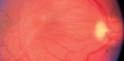
Macular Epiretinal Membrane
 You or someone you know may have been diagnosed as having an epiretinal membrane. The name of this condition has many synonyms, including macular pucker, preretinal fibrosis, surface wrinkle retinopathy, as well as others. This disorder usually causes blurred and distorted vision. We have prepared the following explanation to help you understand this condition better.
Diagnosis of epiretinal membranes
Your ophthalmologist can detect an epiretinal membrane by examining your retina. Sometimes photographs are taken to monitor the stability or progression of an epiretinal membrane. Photographic tests called optical coherence tomography or fluorescein angiography may be done in order to determine the extent of the damage to the underlying retina and possible causes of the epiretinal membrane.

A fluorescein angiogram is a test where sodium fluorescein dye is injected into the veins of your hand or arm and a series of photographs are taken of your retina. The dye is not x-ray dye, and no x-rays are taken. Rather the dye is a photographic dye and only photographs are taken. The fluorescein angiogram allows the physician to evaluate the blood vessels in the retina as well as the retinal layer and the layer underneath the retina. Patients who undergo a fluorescein angiogram often get a mild yellow discoloration of their skin. The fluorescein dye is eliminated from the patient's body through the urine, which is discolored for up to 24 hours following the test. The test is generally safe; however, rare problems, such as allergies to the medication, can occur. Patients who are allergic to x-ray dye are not necessarily allergic to sodium fluorescein. Optical coherence tomography is a newer test which bounces light waves off the retina to obtain an image of the retina in cross section. Optical coherence tomography uses light waves to image the retina very much like sonar waves are used to image the ocean floor. No dye is injected for optical coherence tomography.
Treatment of epiretinal membrane
The only treatment of epiretinal membranes is surgery to remove the membrane. If your symptoms are mild, surgery is usually not necessary. Strengthening your bifocals or using a magnifier may improve your near vision, if both eyes are involved. If your symptoms are significant, then an operation called a vitrectomy is performed to remove the membrane. This surgery is usually performed as an outpatient procedure with a local sedation form of anesthesia. During the surgery, the retina specialist uses tiny instruments to remove the vitreous jelly from the central cavity of the eye and subsequently remove the membrane which is wrinkling the macula. Usually the macula flattens out and the symptoms slowly improve. The majority of patients get improvement of vision following the operation. Vision does not usually return all the way to normal, however, and you may be left with some distortion in your vision, as well as reduced acuity.
